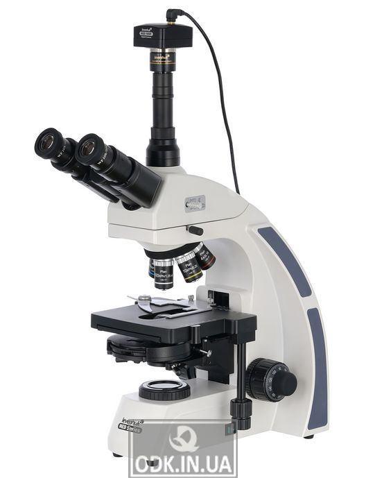
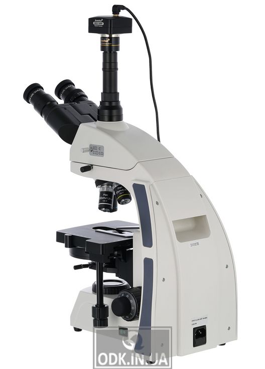
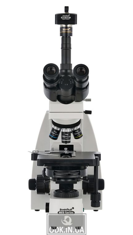
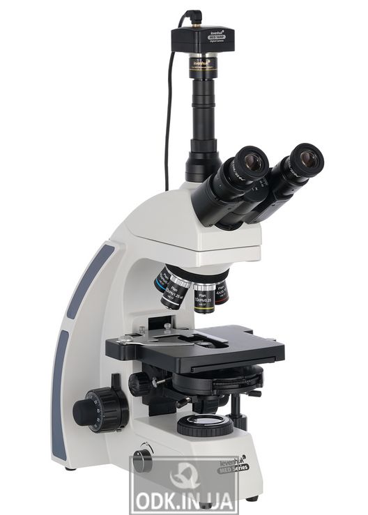
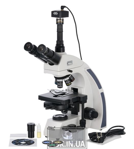
The Levenhuk MED D45T trinocular microscope with a 16-megapixel camera is an opportunity to make visual observations and at the same time record research in photo or video format. The microscope is ideal for professional work in the laboratory, creating digital archives, conducting complex microbiological research. It allows the use of phase-contrast microscopy, light and dark field methods, oil immersion and Keller illumination. The possibilities of the microscope will be evaluated by specialists of research centers, medical institutions, universities.
Advantages of the phase-contrast method The method increases the contrast and sharpness of translucent and transparent samples to a level that in the classical study can be achieved only by staining. Therefore, phase-contrast microscopy is indispensable for living samples in their natural form, because any staining inevitably leads to their death. The method can be used for the analysis of drinking water, parasitological, cytological, hematological and other types of research. class. This system includes Infinity PlanAchromat lenses and allows you to get clear, high-contrast images with a high degree of flatness. One of the most important features of an infinite optical system is that it allows you to establish the optical path of the microscope between -what additional components. These include polarizers and epi-fluorescent illuminators. As a result, the modular design principle and ease of use make the Levenhuk MED 45 optimal microscope for use in various types of microscopy and work in hematology, histology, microbiology and other laboratories.
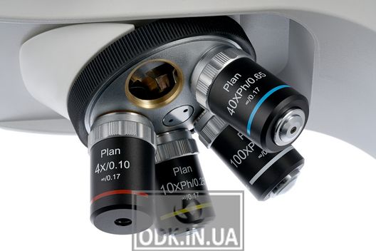
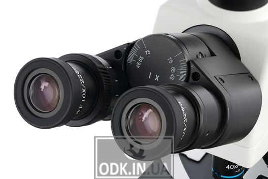
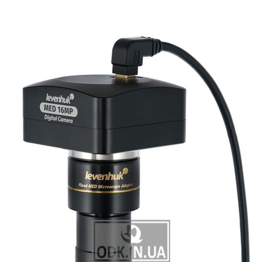
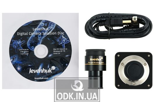
Visual observations without distortion removes chromatism and reduces other distortions. The transmitted image is clear, contrasting and clear. 10-fold glasses give a good overview and allow you to adjust your diopter. The trinocular nozzle is divided into a visual part and a tube for mounting a vertical camera. It rotates 360 °, which is convenient for group research. And a small 30-degree tilt of the visual unit facilitates long-term work with the microscope - the muscles of the neck and shoulder girdle are less tense. Up to five lenses can be installed in the turret device, four are included in the delivery set - these are phase plans of the chromatic model. 40x and 100x lenses have spring frames, and a 100x lens can be used for oil immersion. The optics are made of antifungal glass, have a rough and precise adjustment of sharpness. It transmits the image from the microscope lens in real time, allows you to record videos and take photos. High quality recordings and photos allow them to be used in professional activities. It also comes with a disk with an image processing program.
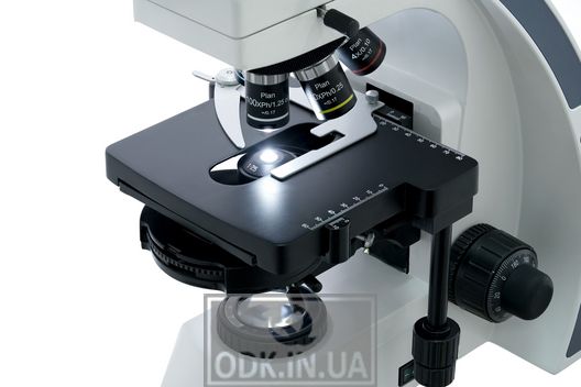
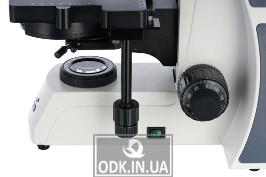
The digital camera makes laboratory work easy and convenient, as it allows you to conduct research without looking at glasses, but on a computer screen, which significantly reduces the load on vision . With the help of the software you can adjust the size, brightness and contrast of the image, change the shutter speed, calibrate the camera with lenses, measure samples or individual structures (several units are available). in two directions. The drug helps to accurately identify samples under the lens of a microscope. The bright 5-watt LED illuminates the micropreparations from below, while the light passes through a phase capacitor ("dry"). There is a filter holder. The brightness of the backlight is adjustable.
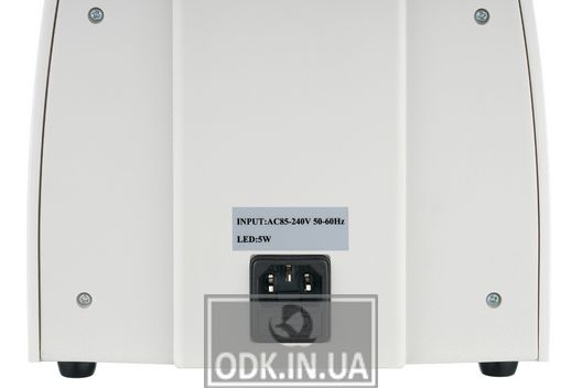
Backlight requires AC connection.
Features:
Trinocular microscope with magnification from 40 to 1000 times.
Phase plans chromatic lenses, adjusted to infinity
Wide-field glasses with diopter correction
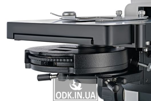
Phase condenser with dark field
LED backlight with brightness adjustment
Adjustable Keller lighting
Digital camera 16 Mpix included
Supplied with: base
Trinocular 360 ° rotary nozzle
Phase planochromatic lenses, infinity-adjusted: 4x, 10x, 40xs, 100xs (oil) with antifungal coating
Wide-field glasses: WF10x / 22 mm with antifungal 2 pcs.)
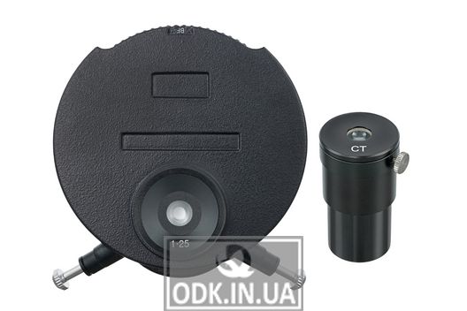
Phase contrast device (dark field)
Filters: blue,
green, yellow
Bottle with immersion oil
Fuse (2 pcs.)
Mains cord for microscope
Dust cover
Digital camera 16 MP x
Camera adapter
USB cable for connecting and powering the camera
Software and driver CD
Operating instructions and warranty card
Attention! Remember that the mains voltage in most European countries is 220-240 V.
narrow voltage standard, you need to plug it into the socket only through the appropriate converter (voltage converter).
This can be seen under a microscope:
The photo shows slides from the Levenhuk micropreparation kits.
The Levenhuk MED D45T microscope is compatible with Levenhuk digital cameras (cameras purchased separately). Levenhuk cameras are installed in the eyepiece tube instead of the eyepiece. The microscope is also compatible with any other digital microscope camera